iTA VIEW is a viewer for DICOM and DICOM RT objects developed for iPad (iPad 3rd generation or later, iOS 6 or later) and dedicated to radiotherapy.
iTA VIEW CANNOT be used as a medical device for primary diagnosis.
With iTA VIEW you can connect to any DICOM node, set filters on "Patient Name" and "Patient ID" and download Studies with the associated Series in your local database. Studies can also be imported into the local database via iTunes File Sharing.
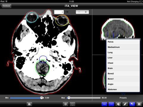
With iTA VIEW you can display CT, MR, PT and 3DUS images and DICOM RT objects, such as RT Images, set of structures, dose distributions and DVHs. The original images (axial view) and the reconstructed planes (sagittal and coronal views) are visualized. The resolution of the reconstructed views is chosen by the user. The Regions of Interest (ROI) contours and dose distributions are displayed on the three views.
iTA VIEW calculates the dose-volume histograms (DVH) and displays the results for the selected ROIs. The DVHs are shown both in absolute and relative dose; selecting a point on the DVH, its dose-volume values are displayed.
iTA VIEW is equipped with tools for drawing and writing that can be used to add comments, lines, arrows and points to the axial views. The pictures with comments and notes are added to the PDF report that is created by the application and which also contains basic information about the patient and the treatment plan and DVHs. Also pictures (patient, positioning system, etc.) and information encoded in barcodes can be added to the PDF report.
- Query/retrieve from all DICOM servers (C-Get, C-Move).
- Import in local database of studies via iTunes File Sharing.
- Search and show Studies in local database (with the free version only two Studies at a time can be stored in the local database).
- Visualization of DICOM images (CT, PT, MR, 3DUS), DICOM RT Images and DICOM RT Objects (ROIs, POIs and DOSEs).
- Traditional 2D visualization: axial, sagittal and coronal views.
- Visualization of ROIs from DICOM RT Structures.
- Zoom and Pan.
- Contrast and intensity adjustment.
- 2D dose visualization from DICOM RT Dose.
- Custom isodoses visualization.
- DVH calculation and visualization (available in the full version).
- Tools for freehand drawing and writing notes on images.
- Take and add pictures (patient, positioning system, etc.) to the PDF report.
- Scan a barcode (or use a photo) and add the encoded information to the PDF report.
- Send reports by email (in PDF format) or to a DICOM node (as DICOM, modality DOC) with selected images, DVH(s), information about the patient and the treatment plan, pictures and barcode information (available in the full version).


